
Fig. 9.
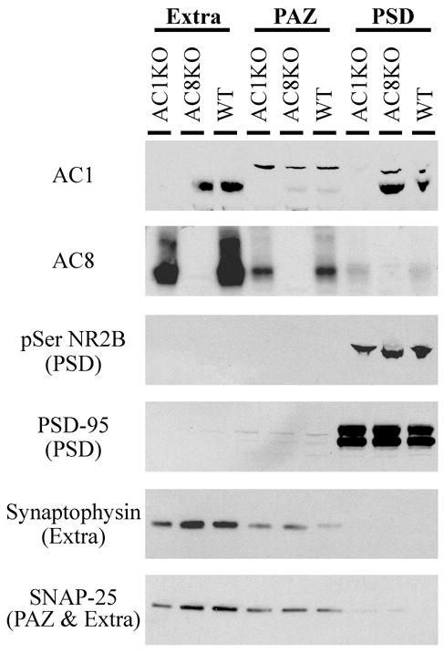
Protein immunoblot analysis of AC1 and AC8 in synaptosome fractions reveals distinct patterns of subcellular distribution. Intense AC1 immunoreactivity is detected in extrasynaptic membrane (extra) and postsynaptic density (PSD) fractions, with minimal expression in the presynaptic active zone (PAZ). In contrast, AC8 is localized to extrasynaptic membrane and PAZ fractions, with minimal expression in the PSD. The extrasynaptic fraction includes both pre- and postsynaptic membranes. Appropriate enrichment of fractions is confirmed with markers of the PSD (pSer NR2B and PSD-95), the extrasynaptic pool (synaptophysin and SNAP-25), and the PAZ (SNAP-25).