
Fig. 2.
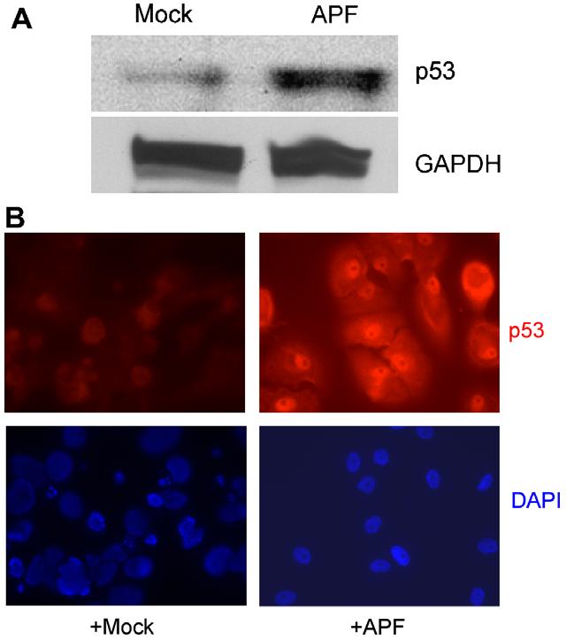
Increased p53 protein expression in response to APF. (A) Whole cell lysates were prepared from urothelial cells treated with 10 ng/ml APF or Mock APF for 7 d. Western blot analysis was performed using anti-p53 and GAPDH Abs. (B) Cells were stained with anti-p53 Ab (red), and nuclei were counterstained with DAPI (blue).