
Figure 3. Changes of suture width in histological sections.
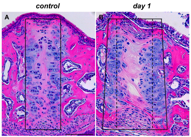
Hematoxylin and eosin staining of frontal sections of midpalatal sutures of control (A) and expansion (B) animals. In control sections, the suture area was contained within a rectangular frame (A); in expansion sections, the suture area was widened and contained within a trapezoidal frame (compare the control width indicated by a stippled line with the expanded width indicated by a solid line in B). The two images, as well as all subsequent images, are oriented with nasal side up and oral side down. Scale bar (A): 100μm.