
Figure 2.
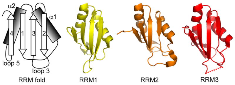
RRMs 1, 2 and 3 in Prp24-N123 crystal structure. The RRMs adopt canonical folds. The schematic at left shows the canonical β–α– β– β–α– β RRM fold, with the β-strands and α-helices numbered in order from the N-terminus. The backbone structure of RRMs 1–3 is shown at right. The dotted line indicates a disordered region in loop 3 of RRM 3.