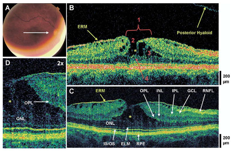
Figure 1.

Lamellar hole and typical epiretinal membrane (ERM). The patient is a 69-year-old woman who presented with a best-corrected visual acuity of 20/40 in the right eye. Dilated fundus examination revealed an ERM in the macula, with a small sharply-circumscribed red lesion at the fovea, which was believed to be a pseudohole. A, Fundus photograph depicting lamellar hole and the direction of optical coherence tomographic (OCT) scans. B, Stratus OCT image demonstrates all 4 criteria for the diagnosis of a lamellar hole, which are labeled 1, an irregular foveal contour; 2, break in the inner fovea; 3, separation of the inner from the outer foveal retinal layers; and 4, absence of a full thickness foveal defect with intact foveal photoreceptors. The posterior hyaloid is detached from the macula, and an ERM of typical appearance is visible. C, Ultrahigh-resolution optical coherence tomographic (UHR OCT) image, which also shows intraretinal separation occurring between the outer plexiform layer (OPL) and the outer nuclear layer (ONL). The foveal photoreceptor layers are intact below the area of foveal dehiscence (i.e., the ONL, the inner/outer segment junction [IS/OS], and the external limiting membrane [ELM]). Other retinal layers and the retinal pigment epithelium (RPE) are labeled as follows: retinal nerve fiber layer (RNFL), ganglion cell layer (GCL), inner plexiform layer (IPL), and inner nuclear layer (INL). D, Magnification (×2) of UHR OCT image shows strands of tissue spanning between the separated ONL and OPL (yellow asterisks), and the intraretinal split.
