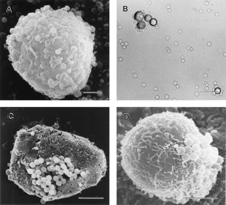
Figure 1.

Binding of MSH beads with melanoma cells. (A) Scanning electron micrograph showing MSH microspheres bound to C-8161 human amelanotic melanoma cells. (Bar = 5 μm.) (B) Light microscopic visualization of specific binding of B16 mouse melanoma cells to MSH macrospheres. (Bar = 100 μm.) (C) Clustering of microspheres observed during binding to B-16 mouse melanoma cells. (Bar = 5 μm.) (D) Human amelanotic melanoma cells (VS). (Bar = 5 μm.)
