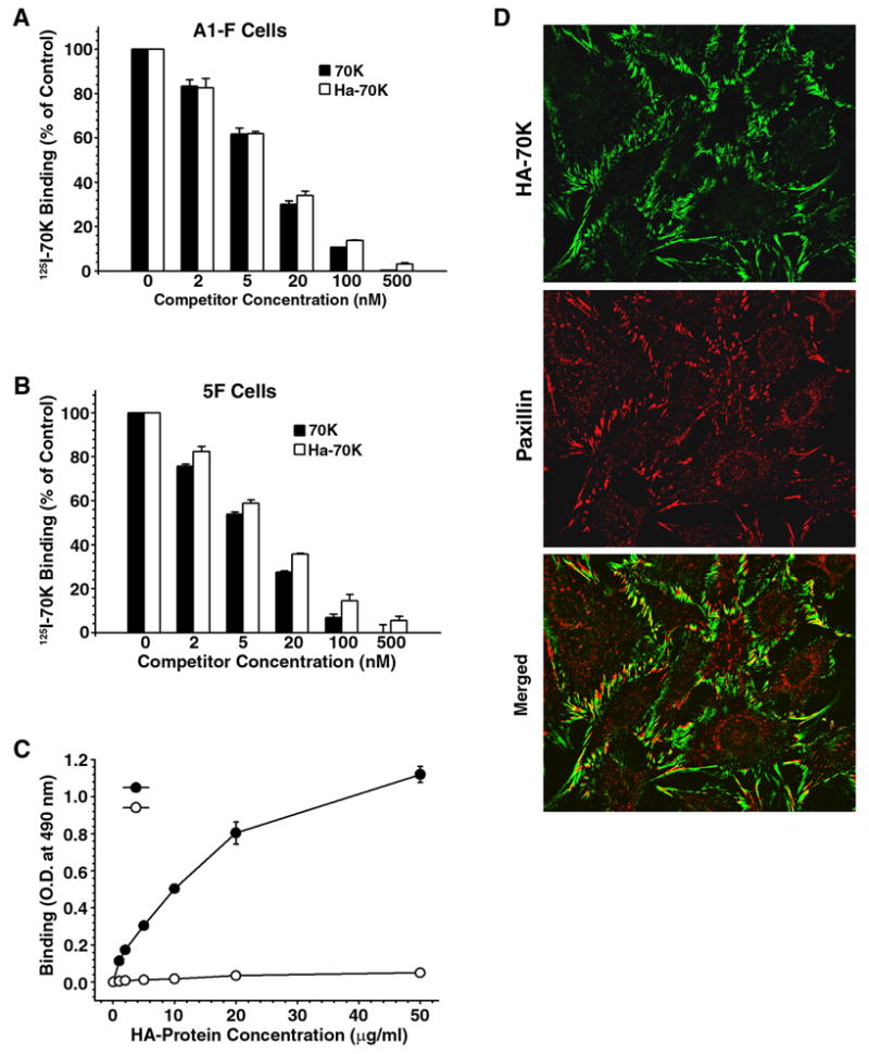
Fig. 1.

Binding and localization of HA-70K to adherent fibroblasts. Human fibroblasts, A1-F (Panel A) and FN-null mouse fibroblasts, 5F cells (Panel B–D) suspended in DMEM containing 0.1% heat-inactivated BSA were plated at confluent densities onto FN-coated plates and incubated overnight. A & B - Cell monolayers were incubated with 5 nM 125I-70K fragment and either unlabeled recombinant HA-70K or the 70K cathepsin fragment at the indicated concentration. 125I-70K binding in wells which did not receive any protein competitor were expressed as 100%. C - Cell monolayers on FN-coated wells were incubated with increasing concentrations of HA-70K or HA-40K. Bound HA-proteins were determined by ELISA as described under Experimental Procedures. (D) 5F cells on FN-coated coverslips were incubated with HA-70K. Cell layers were subjected to dual immunofluorescence staining: HA-70 kDa was visualized using Alexa-488 (green); paxillin was stained with Alexa-594 (red).
