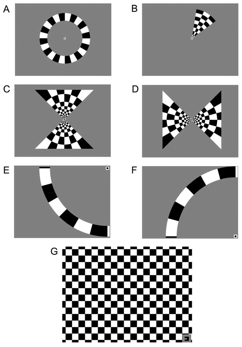
Figure 1. Visual Stimuli for FMRI Experiments.

Several stimuli (A–F) were used to obtain retinotopic maps of the visual world on the flattened cortex of the patients. Patients fixated on a stationary target while contrast-reversing checkerboard patterns (100% contrast; 8 Hz flicker) were presented in the periphery. All stimuli were presented for 6 cycles of 40 s each. A: Expanding rings continually expanded outward from the fixation point each cycle. B: Rotating wedges revolved 360° clockwise around the fixation point each cycle. C–D: Meridian-mapping stimuli. The horizontal and vertical meridians were stimulated using “hourglass” and “bow tie” shaped checkerboard patterns that were alternated every 1/2 cycle. E–F: 16° isopter stimuli. A 16° arc subtended the superior or inferior quadrant of the hemifield containing the scotoma. Each arc was presented alternately with a period of no stimulation every 1/2 cycle. G: Scotoma-mapping stimulus. A contrast-reversing checkerboard pattern was presented to the quadrant of visual space with the scotoma. Patients viewed the stimulus through the right or left eye in alternating 1/2 cycles. The fixation target in the corner also served as a cue as to which eye should be open.
