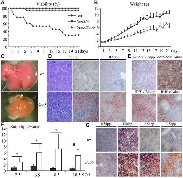
Figure 3.
Growth and liver defects in KitSco5 homozygotes during post-natal development. (A) Viability and (B) body weight curves of Sco5/Sco5 individuals generated from heterozygous Sco5/+ intercrosses (Sco5/Sco5 n = 13; Sco5/+ n = 28; wt n = 16) reveal that mutant homozygotes are strongly affected during post-natal development compared to control littermates. (C) At 7.5 dpc, livers of mutants have a more yellowish color which was associated with the appearance of some white areas (white arrowhead). These alterations were never observed in wild-type (wt) littermates. (D) Hematoxylin and eosin, and Oil-red-O stainings reveal the swelling of hepatocytes with formation of large lipid-containing vesicles in Sco5 homozygotes versus wild-type mouse littermates at 7.5 and 10.5 dpp (magnification ×40). (E) Staining of Sco5/+ individuals at 7.5 dpp shows a relative increase in lipid droplets inside hepatocytes compared to wild-type (D). A similar defect is observed in the liver of 7.5 dpp old mice, surviving adult Sco5 homozygotes and W/W-v compound Kit mutants. (F) The lipid accumulation in the liver was studied by NMR during post-natal development at various ages on wt (n = 5; black) and mutant (n = 9,8,8,3; white) individuals. No statistical analysis was possible at 10.5 dpp because of the death of 6 mutants out of 9 (#), but the lipid/water ratio is still increased in mutant mice, at 10.5 dpp, in coherence with mortality scale and histology. (G) At 0.5 dpp, the Oil-red-O staining of liver sections from Sco5 homozygous and control mice which had not yet suckled, are identical showing no accumulation of lipids. However, after breast feeding, hepatocytes from control newborns appear to stock lipids inside microvesicles at 1.5 dpp, a phenomenon that is not observed at later stages. On the contrary, in mutant mice which have suckled milk from their mother, lipids accumulation is still found in hepatic cells after 1.5 dpp.

