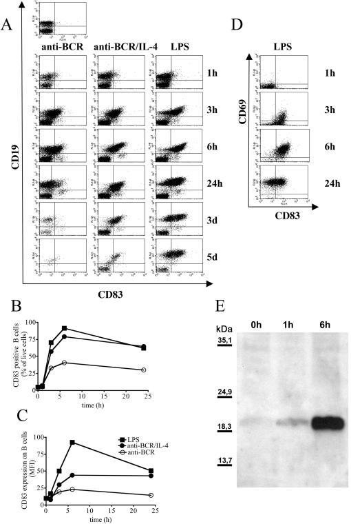
Figure 1. CD83 is upregulated on activated B cells.
C57BL/6 mice derived spleen cells (2×10⁶/ml) were stimulated with anti-BCR (1 µg/ml) and IL-4 (20 ng/ml) or with LPS (10 µg/ml) as indicated in the headline. Cells were triple stained for CD19, CD83 and CD69 at the indicated time points. 1A: Dot blots show all lymphocytes positive for surface expression of CD83 on the x-axis and CD19 expression on the y-axis. 1BC: Graphs show the percentage of CD83 positive B cells (1B) or the mean fluorescence intensity (MFI) of CD83 on B cells (1C) after stimulation with anti-BCR alone (open circle) anti-BCR and IL-4 (closed circle) or with LPS (closed square) in an independent experiment, error bars show SEM of duplicates. 1D: Dot blot shows 2×10⁴ CD19 positive cells derived from LPS activated spleen cells analyzed for CD83 (x-axis) and CD69 (y-axis) surface expression. 1E: 2×10⁶ purified C57BL/6 spleen derived B cells were stimulated with LPS (10 µg/ml). B cells were lysed at the indicated time points, deglycosylated and separated by SDS-PAGE. CD83 was detected by western blot with a polyclonal rabbit anti-CD83 serum. Results are representative for at least three independent experiments.

