
Fig. 10. Effect of rotenone on TH-IR.
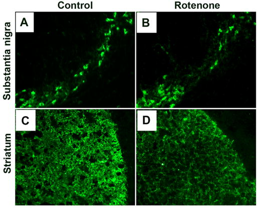
Representative photomicrographs of immunofluorescent staining for TH-IR in coronal brain sections (10 μm) passing through SN and striatal region from control rats (A and C) and rotenone-injected rats (B and D). In control animals, numerous TH-positive cells and intense TH-IR are seen in SNpc (A) and striatum (C), respectively. Rotenone administration caused substantial decrease in TH-IR in both SN and striatum. Images were taken at 200X magnification. (n≥4).