
Fig. 2.
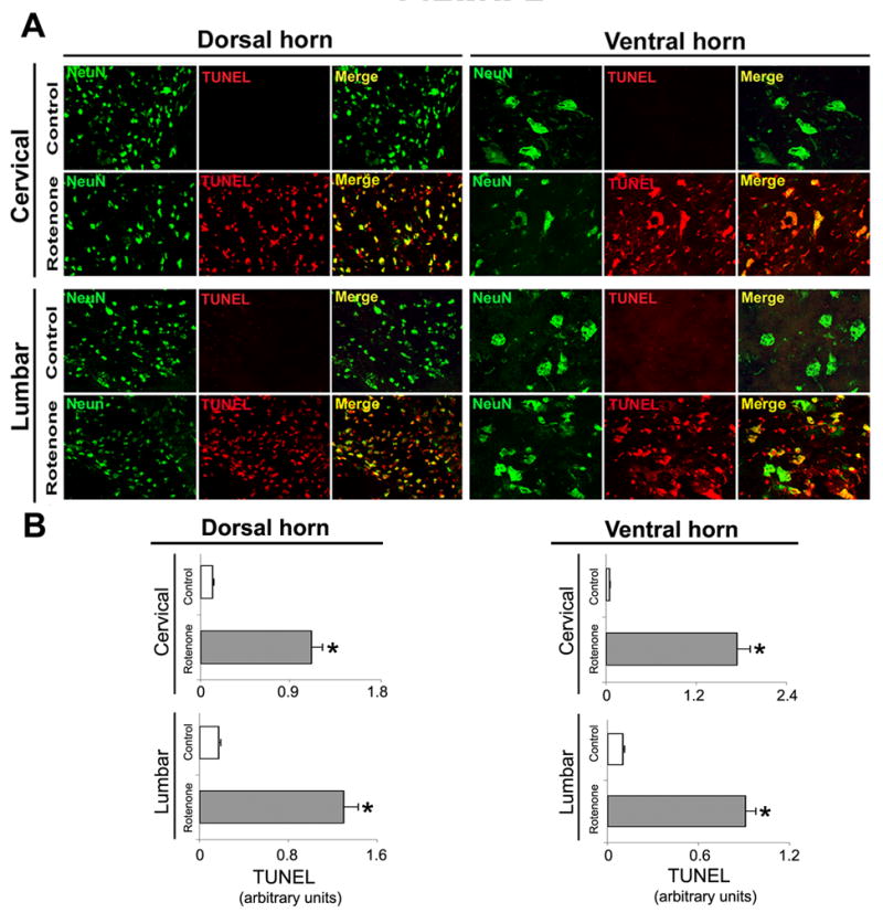
Effect of rotenone on neuronal viability in cervical and lumbar SC. (A) Representative photomicrographs of double immunofluorescent staining for NeuN (green) and TUNEL (red) performed in cervical and lumbar SC coronal sections (5 μm). No TUNEL+ neurons were identified in the sections from control animals (see Merge); however, cell death as evaluated by TUNEL-positive IR in many dorsal and ventral neurons was found in sections from cervical and lumbar SC segments in rotenone-injected animals (yellow, merge). Images taken at 200X magnification (n≥4). (B) The lower panels show semi-quantitative analysis of TUNEL mean fluorescence intensities (MFI) per unit area of neurons. Data show significant differences in the arbitrary units between samples from control animals and after rotenone exposure. *P≤0.05 (n≥ 4).