
Fig. 4.
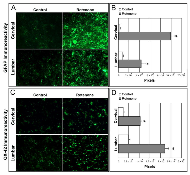
Effect of rotenone on GFAP and OX-42 immunoreactivity in cervical and lumbar SC. In control rat SC sections, there were a few GFAP and OX-42 positive cells. Intense astrogliosis, detected by increased GFAP-IR (A) and microgliosis, detected by OX-42-IR (C) seen in both cervical and lumbar SC slices from rotenone-injected animals. Data analyzed with NIH image 1.63 showed significant differences in the number of pixels in sections from control and rotenone-injected animals (B and D). *P≤0.05 (n≥ 4).