
Fig. 8.
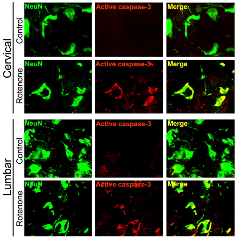
Effect of rotenone on activation of caspase-3 in cervical and lumbar SC. Representative photomicrographs of double immunofluorescent staining for active caspase-3 (red) and NeuN (green), performed in cervical and lumbar SC coronal slices (5 μm). No active caspase-3-IR was found in the sections from control animals. Significant amount of active caspase-3 was observed in many ventral motoneurons in cervical and lumbar SC segments of rotenone-injected animals, evaluated by increased co-localization of active caspase-3-IR and NeuN-IR (Merge indicate co-staining in rotenone panel). Images are taken at 200x magnification (n≥4).