
Figure 5.
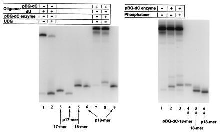
(Left) Denaturing PAGE (20% gels) of 3′-end-labeled fragments obtained from uracil-DNA glycosylase (UDG) activity on a 25-mer oligonucleotide containing dU at the same position as used for pBQ-dC. Lane 2 shows that after UDG treatment the fragment migrates with p17-mer (lane 4) and not with the 17-mer (lane 3). The same procedures for identification of the pBQ-dC endonuclease-treated oligomer (lane 8) did not produce the 17-mer, p17-mer, the 18-mer (lane 5), or p18-mer (lanes 6 and 9). (Right) Size and charge markers to identify the 3′ cleavage product of the pBQ-dC oligomer are in lanes 4–6. Only a synthetic pBQ-dC-18-mer (lane 4) coincided in migration mobility with the pBQ-dC endonuclease-produced 3′ fragment after dephosphorylation (lane 3). This gel shows that it is possible to distinguish between phosphorylated (lane 2) and dephosphorylated (lane 3) cleavage products.