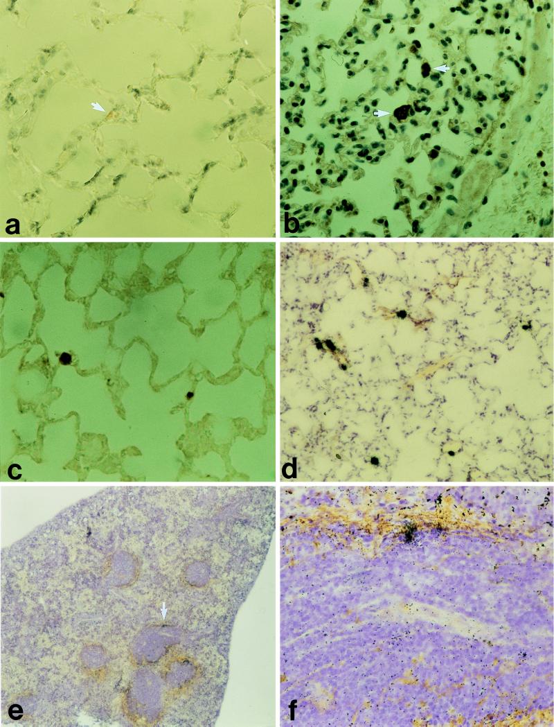
Figure 1.
(a–d): Lung tissue from a mouse given synthetic TTR(115–124) fibrils i.v. Small amyloid deposits (arrows) occasionally were found in vascular lumina stained with Congo red and viewed in polarized light (a) and labeled with antiserum to mouse protein AA and to the synthetic peptide TTR(115–124) (b and c). (d–f) Tissues taken from a mouse that had received 125I-labeled TTR(115–124) fibrils i.v. 22 days before it was killed. (d) Lung tissue with several radioactivity labeled spots corresponding to the minute amyloid deposits. e A section of the spleen and silver grains occur perifollicularly at the amyloid deposits. (f) Close-up of e at the arrow.

