
Figure 4.
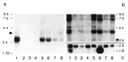
Northern blot analysis of BRCACox in human tissues. A human multiple tissue blot (CLONTECH) containing ≈1 μg of purified mRNA of different tissues was analyzed for the presence of hBRCACox mRNA (a). Lanes: 1, heart; 2, brain; 3, placenta; 4, lung; 5, liver; 6, skeletal muscle; 7, kidney; 8, pancreas. After longer exposures, signals corresponding to BRCACox mRNA became also visible in lanes 2–4 (data not shown). For comparison, in b, the signals obtained for hPALMCox mRNA are shown, while the amount of β-actin mRNA is shown in c. Despite the stringent washing conditions, a very slight cross-reactivity between the mRNAs for hBRCACox and hPALMCox was noticed (marked by solid arrowheads). This is probably due to a stretch of bases with high homology in the middle portion of the mRNA sequences. The migration of standards (expressed in kb) are indicated on both sides (a and b). The values at the right of c correspond to the length (in kb) of the mRNAs for the distinct β-actin isoforms.