
Fig. 6.
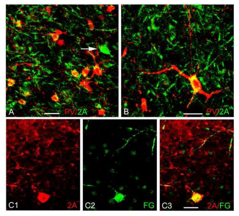
Dual localization of 5-HT2AR immunoreactivity (BD Pharmingen monoclonal antibody) and neuronal markers in the BLa using immunofluorescence confocal laser scanning microscopy. A, B) Dual localization of 5-HT2AR (green) with PV (red). Colocalization is indicated by yellow. Note light to moderate 5-HT2AR immunoreactivity in some of the PV+ neurons. Arrow in A indicates one of the large intensely-stained 5-HT2AR+ nonpyramidal cells that are labeled by injections of fluorogold into the mediodorsal thalamic nucleus (see C1-C3); none of these cells expressed the interneuronal markers PV, SOM or CR. C1-C3) Dual localization of 5-HT2AR (red) and fluorogold (FG; green), with injections of FG into the mediodorsal thalamic nucleus. Note that a large intensely-stained 5-HT2AR+ nonpyramidal cell (lower part of field) is retrogradely labeled by fluorogold. All scale bars = 25 μm.