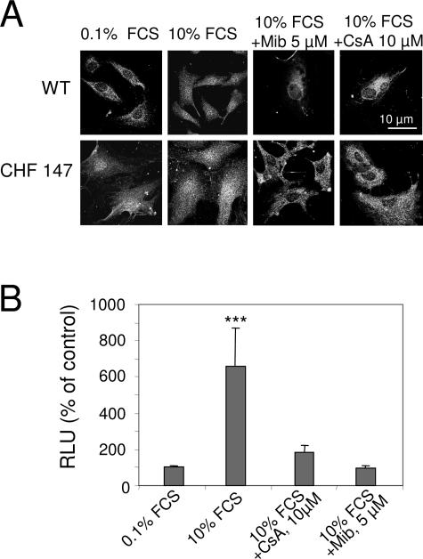
Figure 6.
Involvement of NFAT in VSMC proliferation. A: Confocal immunofluorescence with anti-NFAT showing its cytosolic or nuclear localization in VSMCs. B: Promoter-reporter assay of NFAT transcriptional activity in WT VSMCs. Cells were transfected with NFAT-Luc and cultured for 48 hours without FCS. In A and B, cells were then treated with 10% FCS alone or together with 5 μmol/L mibefradil (Mib) or 10 μmol/L CsA for 5 hours. RLU, relative luminescence units. The bars represent mean ± SEM of three experiments in triplicate. ***P < 0.001 versus control (0.1% FCS).

