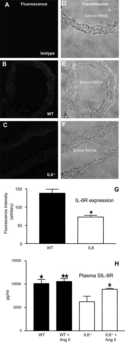
Figure 2.
Aortic IL-6R and plasma sIL-6R is significantly decreased in IL-6−/− mice. Aortic sections from WT and IL-6−/− mice were sectioned and stained for IL-6R. Representative sections are shown for each condition. A–C: IL-6R fluorescence; D–F: corresponding phase-contrast images. G: Mean pixel intensity was determined after fluorescence staining of multiple aortic sections (n = 6, separate aortae, mean ± SEM; ★P < 0.0002, compared with WT controls, unpaired Student’s t-test). H: Effect of Ang II infusion on plasma sIL-6R levels, determined by enzyme-linked immunosorbent assay (n = 6 per group, mean ± SEM; ⋆P < 0.05, IL-6−/− group compared with IL-6−/− Ang II-infused group, ★P < 0.02, ★★P < 0.005, IL-6 −/− group compared with WT and WT Ang II-infused group; unpaired Student’s t-test).

