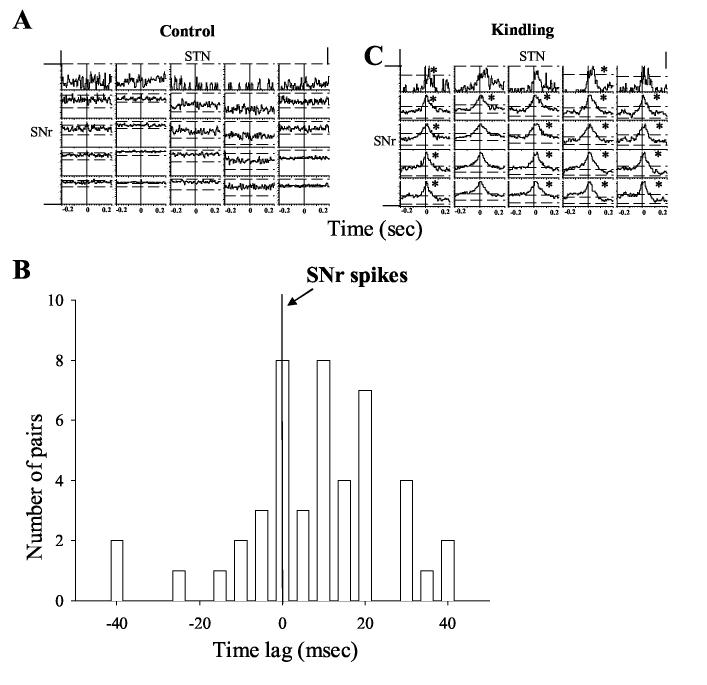
Figure 7.
Temporal relationship between STN and SNr neurons during amygdala kindled seizures. A) Cross-correlogram plots from selected STN and SNr neurons in the kindling side before (left panel) and during amygdala kindled seizures (right panel). SNr neurons were used as reference neurons. Synchronization occurred in these neurons only during amygdala kindled seizure (displayed in the right panel). Most STN neurons lagged behind SNr neurons in synchronized firing (Marked by * signs). B) Distribution of time lags between synchronized firing of STN and SNr neurons (SNr neurons were reference neurons). Most of the STN spike peaks followed SNr spikes with time lags up to 40 ms.

