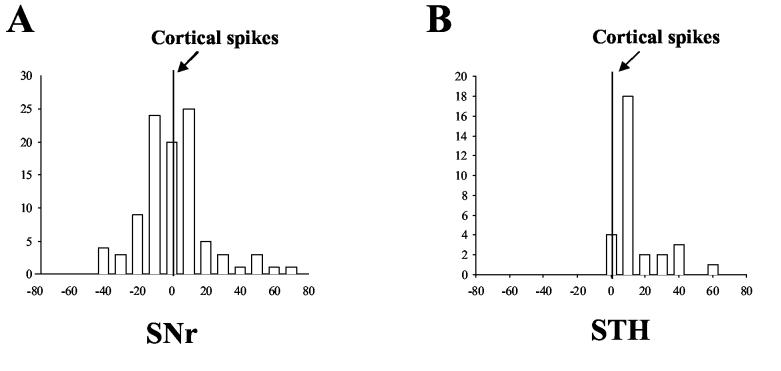
Figure 8.
Temporal relationship of correlated firing between Ctx and basal ganglia neurons in the kindling side during amygdala kindled seizures. A) Distribution of correlated spikes between cortical and SNr neurons using cortical neurons as a reference. Synchronized SNr spikes were distributed evenly around the cortical spikes at the 0 s reference point with an average time lag of −1.4 ±1.5 ms. B) Distribution of correlated spikes between cortical and STN neurons. All correlated STN spikes were located on the plus side of the time lag with a peak at 10 ms.

