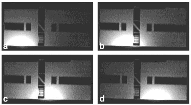
FIG. 10.

a–d: Axial images of each separate strip of the four-strip TW-PSA during a parallel detection from a GE Medical Systems standard phantom containing rectangular signal voids. The images, which correspond to the sensitivity profiles of each strip, show no mutual interference, even in regions where the elements have overlapping sensitivity. FOV = 28 cm.
