
Figure 3.
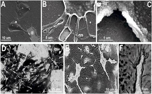

(A) SEM image of cell-templated PEDOT electrode coating prepared by enzymatic and mechanical removal of cells and cell material which reveals cell-shaped holes (@). This image also shows some cells that are completely covered and trapped in the PEDOT matrix (*). (B) Neurite-templated tunnels and crevasses are created in the PEDOT matrix by removing cells after polymerization. (C) Higher magnification image further reveals the details of the neurite tunnels and the polymer surface shows a “negative” image of the neurite membrane surface. (D) and (E) Optical images of neuron-templated PEDOT film on Au/Pd electrode shows detailed PEDOT “template” of where neurons where adhered to the substrate during polymerization. (F) PEDOT uses extracellular matrix and cell membranes as scaffolds for polymerization making possible visualization of micro and nano-filopodia allowing formation of an intimate interface between the neuron and the conductive matrix. (G) Atomic force microscopy (AFM) image of a neuron-templated PEDOT.