
Figure 7.
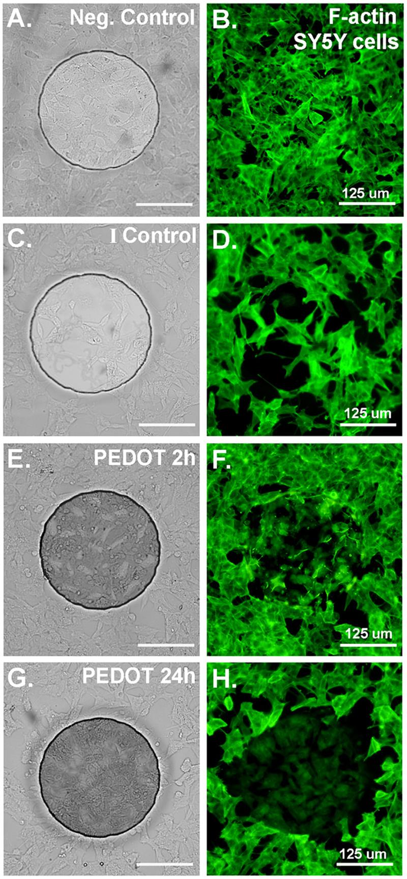
(A) Bright-field image of SY5Y cells cultured on ABP electrode (negative control; no PEDOT, not exposed to current). (B) SY5Y cells cultured on ABP electrode (negative control; no PEDOT, not exposed to current) stained for F-actin with Phalloidin-Oregon Green (green fluorescence). (C) & (D) Cells exposed to current only (no PEDOT), fixed and stained 24h after current exposure shows some morphological alterations but F-actin staining remains robust. (E) & (F) Cells 2h after PEDOT polymerization begin to show abnormal cytoskeletal staining and loss of F-actin stress fibers. (G) & (H) Cells 24h after PEDOT polymerization show a nearly complete loss of F-actin stress fibers and a decrease in the intensity of F-actin staining overall.