
Figure 2.
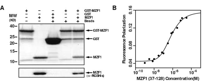
Residues 37-128 of MZF1 contain a SCAN domain that self-associates.
(a) GST-MZF1(37-128) binds MZF1(37-128). Sample composition is indicated above each lane. Proteins were detected by Coomassie stain (upper panel), and the MZF1 SCAN domain was detected by Western blot with a monoclonal antibody specific for the α-RGSHis epitope. Control lanes show that His-MZF1 does not bind non-specifically to GST or glutathione-agarose beads. (b) Tryptophan fluorescence polarization values for MZF1 SCAN domain are plotted as a function of protein concentration. Nonlinear fitting (solid line) yielded an equilibrium dissociation constant for the MZF1 scan homodimer of 600 ± 100 nM and FP values of 0.045 and 0.159 for pure monomer and dimer, respectively.