
Figure 7.
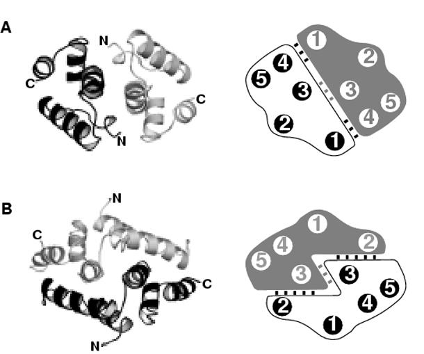
Topology of the SCAN dimer interfaces.
Representative structures and schematic representations of the alternative (a) and domain-swapped (b) dimer interfaces showing the helical interactions. (a) The alternative dimer interface is represented by the HIV capsid C-terminal domain (PDB entry 1a43). One half to the dimer is colored in gray and the opposing monomer is colored in black. (b) The domain-swapped interface is represented by the MZF1 SCAN domain (PDB entry 2fi2).