
Fig. 3.
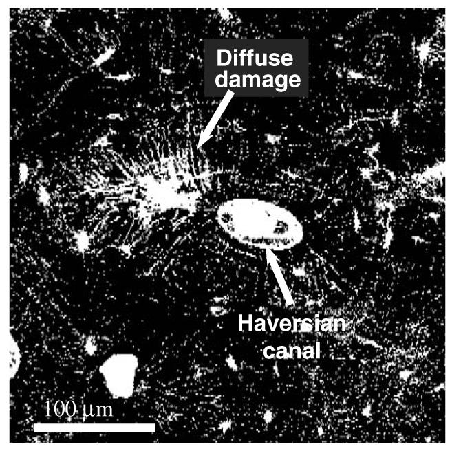
A fluorescence microphotograph (laser confocal microscopy) of a diffuse damage in a transverse cross section of a human cadaveric femur: the bone sample was stained with basic fuchsin following the procedure provided by Burr and Stafford (1990). The fluorescence microphotograph indicates that the diffuse damage comprises a large amount of submicron level defects (nano crack arrays).