
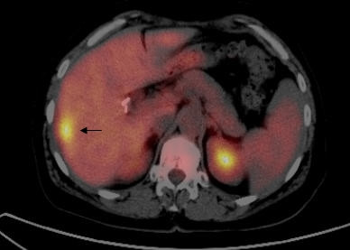
Figure 2.
PET/CT fusion image on transaxial section demonstrates a hypermetabolic lesion (SUVmax : 8.2, Patient number 14) in the segment 6 of the liver (arrow) which could not be detected by GP. Final pathology was consistent with CRC metastasis.
Official websites use .gov
A
.gov website belongs to an official
government organization in the United States.
Secure .gov websites use HTTPS
A lock (
) or https:// means you've safely
connected to the .gov website. Share sensitive
information only on official, secure websites.
PET/CT fusion image on transaxial section demonstrates a hypermetabolic lesion (SUVmax : 8.2, Patient number 14) in the segment 6 of the liver (arrow) which could not be detected by GP. Final pathology was consistent with CRC metastasis.