
Figure 2.
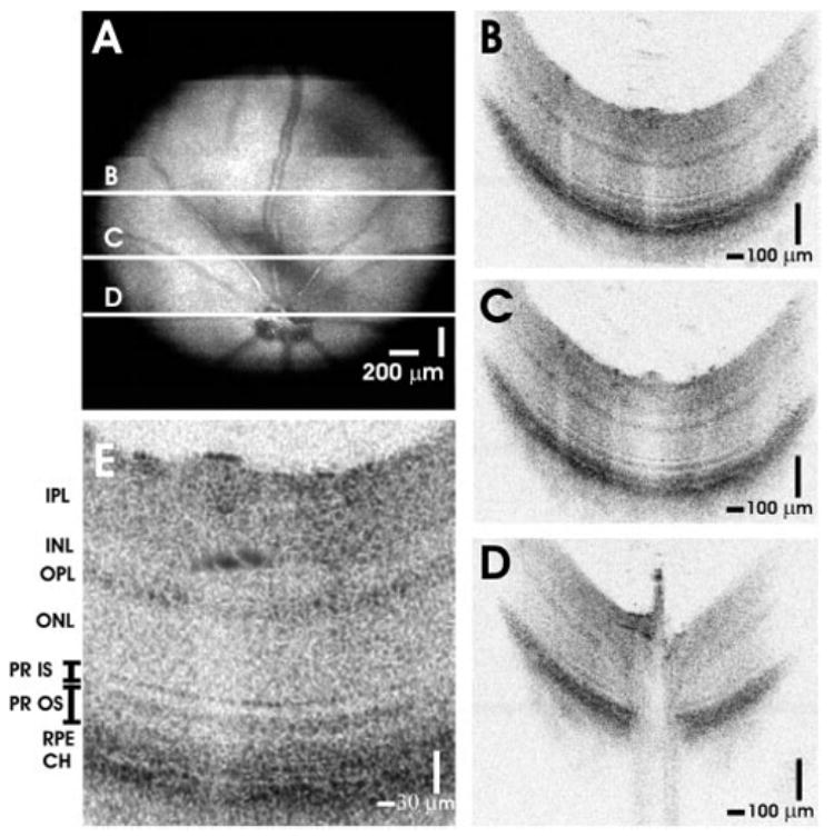
High-speed, ultrahigh-resolution OCT images from a normal C57BL6 mouse. (A) An OCT fundus image, created by axial summation of 3D-OCT data consisting of 256 images of 512 axial scans each, is shown. High-definition OCT images (B–D) with 2048 axial scans may be registered to the OCT fundus image. (E) Clear visualization of major intraretinal layers is enabled by OCT, as shown in the cropped, enlarged OCT image with ~600 axial scans.