
Figure 4.
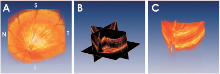
Using 3D-OCT data, visualization techniques such as volumetric rendering are possible. (A) A rendering of a normal Long-Evans rat retina. (B) It is possible to create virtual slices through 3D-OCT data and view images along arbitrary planes. Cut-away renderings can simultaneously show intraretinal structure and retinal topography (C).