
Figure 2.
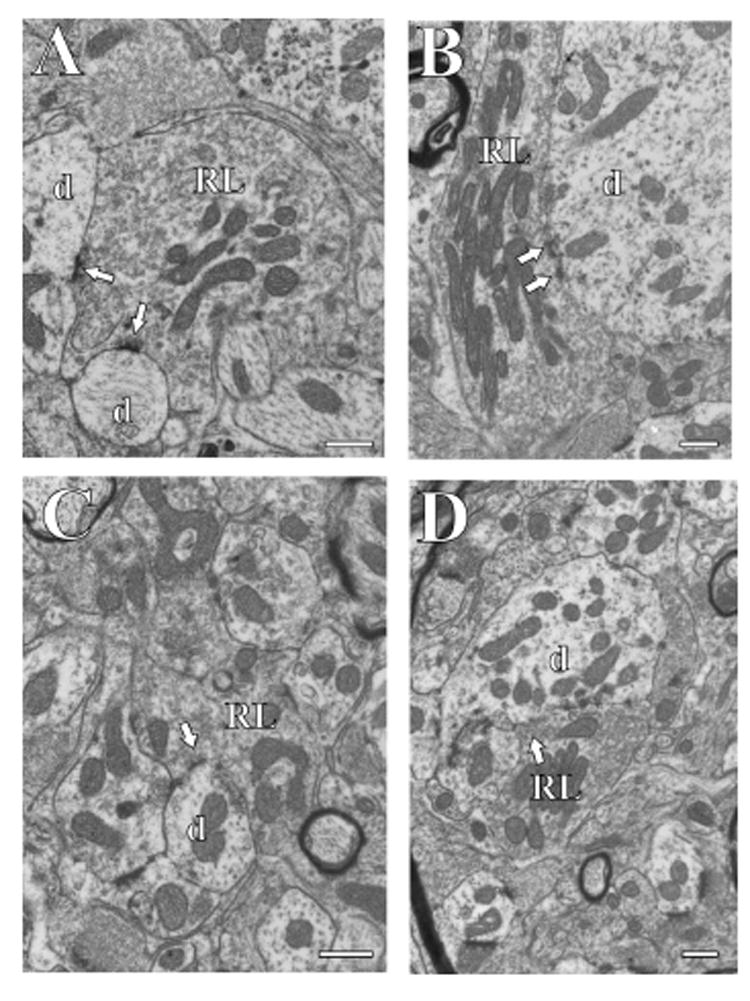
Electron micrographs of RL terminals (RL) in different thalamic nuclei. The arrows point to synapses, and postsynaptic dendrites (d) are also shown. The scale bar in each panel is 0.5 μm. A: Posterior nucleus. B: Ventral posterior nucleus. C: Ventral portion of the medial geniculate nucleus. D: Medial portion of the medial geniculate nucleus.