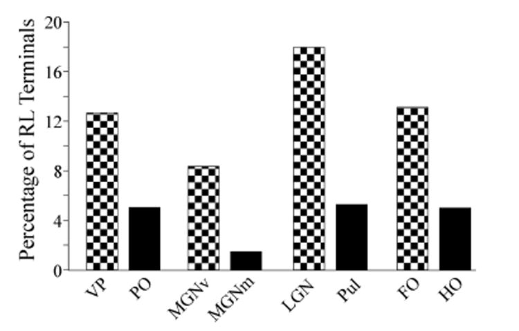
Figure 5.

Relative percentage of synapses from RL terminals in various thalamic nuclei. In each case, the first order nuclei are indicated by hatched bars, and the higher order nuclei by solid bars. data for the left 4 thalamic nuclei are from the present paper; data from the LGN are from Van Horn et al. (2000) and from the Pul are from Wang et al. (2002a). The averages for the right hand pair of bars were computed by averaging the RL percentages from each of the other thalamic nuclei so that different sample sizes between nuclei does not distort the average. Abbreviations: FO, first order thalamic nucleus; LGN, lateral geniculate nucleus, HO, higher order thalamic nucleus; MGNm, magnocellular portion of the medial geniculate nucleus; MGNv, ventral portion of the medial geniculate nucleus; PO, the posterior nucleus; Pul, pulvinar, VP, ventral posterior nucleus.
