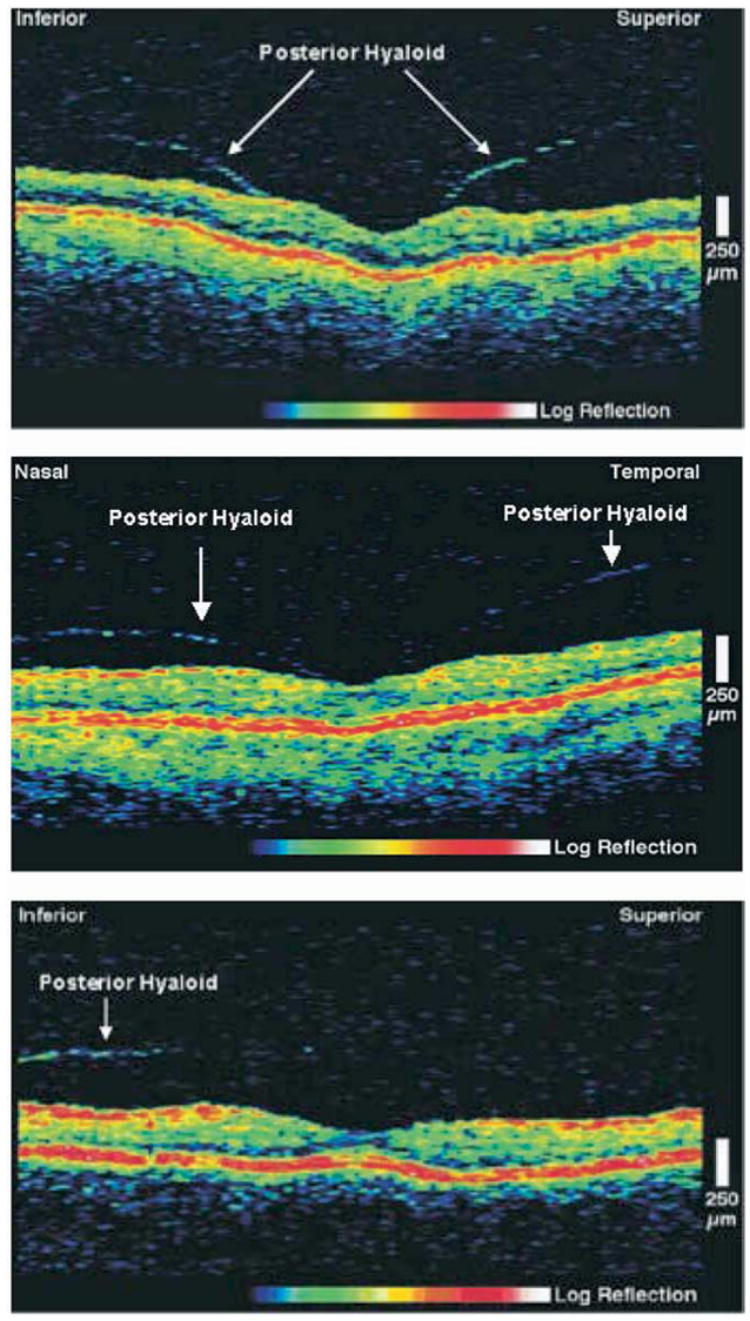
Figure 1.

Optical coherence tomography of fellow eyes with abnormalities at the vitreoretinal interface, classified according to the morphology and severity of the signal from the visible posterior hyaloid. Top, Severe case: prominent insertion of posterior hyaloid on both sides (superiorly and inferiorly) of the perifoveal region. Middle, Moderate case: prominent insertion of posterior hyaloid on only one side (nasal) of the perifoveal region. There is no distinct point of insertion on the other side (temporal). Bottom, Mild case: a preretinal signal corresponding to the posterior hyaloid is visible inferiorly, but there is no clear point of insertion. No posterior vitreous detachment was found on clinical examination.
