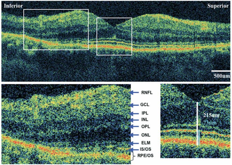
FIGURE 2.

Ultra-high resolution optical coherence tomography (UHR-OCT) of eye with retinitis pigmentosa (RP) and normal visual acuity. UHR-OCT macular images from the left eye of case 6. Visual acuity is 20/20. (Top) A 6-mm vertical scan. (Bottom right) 2× magnification of the extrafoveal macula. Note thinning of the outer nuclear layer and disappearance of the external limiting membrane and photoreceptor inner/outer segment junction. Retinal layers are labeled. (Bottom left) 2× magnification of the fovea, showing central foveal thickness (CFT) (white) and foveal outer segment/pigment epithelium thickness (FOSPET) (blue) measurements. Both thickness measurements are within the normal range.
