
Figure 3.
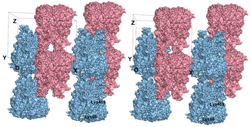
Crystal packing in the P321 unit cell of CELO hexon. The stereoview is almost along the crystallographic twofold axis. There are two hexon trimers in the unit cell (pink and blue), with their molecular axes aligned along the crystallographic threefold axes. These form anti-parallel columns of hexons throughout the crystal. The crystal has alternating layers of hexons with the same color that interlock with each other. Lys65 and Lys459, which lie on the surface of the channel through the crystal, are highlighted in gold and labeled. The CELO hexon model was produced by JACKAL. All residues are included, except those at the tip of the DE1 loop (residues 135–169) that are disordered in the AdH5 and AdH2 hexon crystal structures and so were unconstrained in the molecular modeling. Figure produced with PyMol (DeLano, 2002).