
Figure 1. Retinal function loss following intravitreal injection of wild type and quorum sensing-defective Bacillus cereus and supernatants.
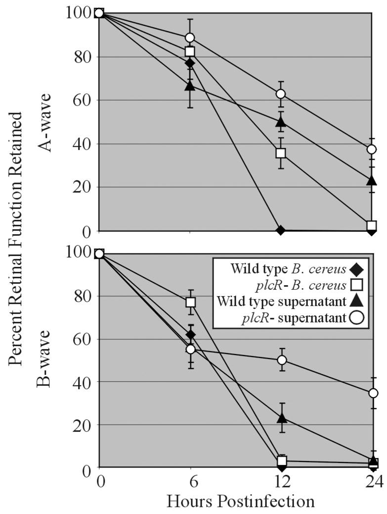
Mouse eyes were injected into the mid-vitreous with approximately 100 CFU/100 μl of wild type (◆) or plcR- (□) B. cereus or 100 μl of a 10-hour sterile filtered culture supernatant of wild type (▲) or plcR- (○) B. cereus. Data at each time point are presented as the percentages of A-wave or B-wave amplitude of retinal function retained compared to baseline and sham-injected controls. Data was averaged for each group and compared using the Student t test. Values and error bars represent means ± standard errors of the means for four or more eyes per group.