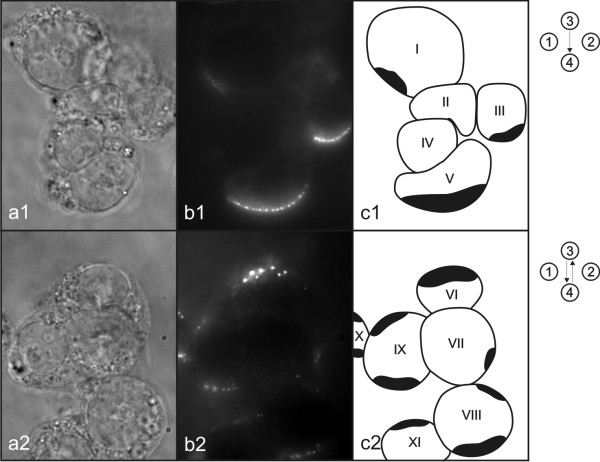
Figure 8.
Shading effect. Photos of phase contrast (a) and fluorescence (b) images were taken under inverted fluorescence microscope. Symbolic picture (c) was made for better representation of the observed shading effect. Drawn shapes represent cells and black areas represent regions of permeabilized membrane where DNA interacts with cell membrane. Symbols on the right represent electric field protocol used.

