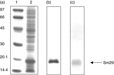
Fig. 3.
SDS-PAGE and Western blot analysis of the recombinant Sm29–6xHIS fusion protein. (a) Coomassie blue stained SDS-12% PAGE profile of induced E.coli expressing the pET21a-Sm29 construct; 1, ladder (kD); 2, E. coli lysate expressing Sm29. (b) Comassie blue stained SDS-12% PAGE profile of the purified Sm29–6xHIS fusion protein. (c) Western blot analysis of the purified protein using anti6xHIS antibody. Arrow indicates the rSm29.

