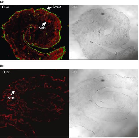
Fig. 4.
Immunolocalization of Sm29 on S. mansoni tegument. Fluorescence confocal microscopy images (Fluor) and corresponding differential interface contrast (DIC) images of male adult worm of S. mansoni are shown. Polyclonal anti-Sm29 and secondary antibody coupled to FITC were used for fluorescence detection of Sm29 on male adult worm sections. Serum from naive mice was used as negative control. Flavoidin and Rodamin were used for the actin localization. The Sm29 localization is represented in (a) and the negative control is shown in (b).

