
Fig. 3.
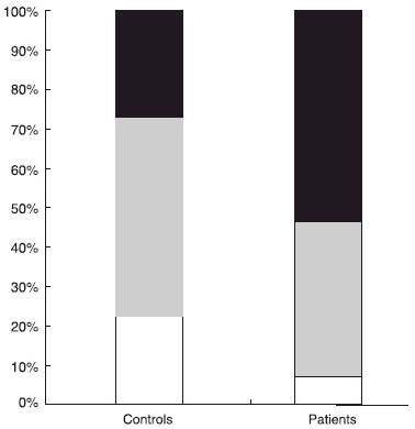
Phenotype distribution of membrane-proteinase 3 (mPR3) on neutrophils. Individuals divided into three predefined groups, high (black)/intermediate (grey)/low (white), according to their percentage of mPR3+ neutrophils [6]. The distribution of these three groups is shown in 58 patients with anti-neutrophil cytoplasm antibodies-associated systemic vasculitis compared to 107 healthy blood donors.