
Fig. 3.
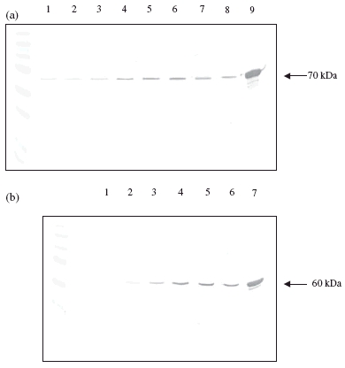
Western blots showing the presence of heat shock protein (Hsp)60 and Hsp70 in peripheral blood mononuclear cells media and cell extracts following 24 h treatment with Hsp60, Hsp70, GroEL and lipopolysaccharide (LPS). (a) Hsp70 in media: lane 1, control; lane 2, Hsp60 (10 µg/ml); lane 3, LPS (10 µg/ml); lane 4, GroEL (10 µg/ml). Hsp70 in cells: lane 5, control; lane 6, Hsp60 (10 µg/ml); lane 7, LPS (10 µg/ml); lane 8, GroEL (10 µg/ml); lane 9, Hsp70 standard. (b) Hsp60 in media: lane 1, control; lane 2, LPS (10 µg/ml); lane 3, Hsp70 (10 µg/ml). Hsp60 in cells: lane 4, control; lane 5, LPS (10 µg/ml); lane 6, Hsp70 (10 µg/ml); lane 7, Hsp60 standard.