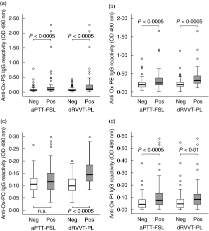
Fig. 3.
IgG reactivity against oxidized phosphatidylserine (a), phosphatidylethanolamine (b), phosphatidylcholine (c) and phosphatidylinositol (d) in the sera of subjects positive (empty bars) or negative (filled bars) for, respectively, activated partial thromboplastin time (aPTT-FSL) or diluted Russell's viper venom test plus phospholipid supplementation (dRVVT-PL) tests. Among the 164 subjects investigated 74 had abnormal aPTT-FSL, while 48 has abnormal dRVVT-PL values. The sera were tested at 1 : 50 dilution in microplate enzyme-linked immunosorbent assay plates coated with the different antigens and revealed with peroxidase-linked goat anti-human IgG anti-serum. The results are expressed as optical density (OD) at 490 nm after subtracting the background reactivity of each serum. Boxes include the values within 25th and 75th percentiles and the horizontal bars represent the medians. Eighty per cent of the values are comprised between the extremities of the vertical bars (10th−90th percentiles). The extreme values are represented by individual points. Statistical significance was evaluated by non-parametric Mann–Whitney U-test.

