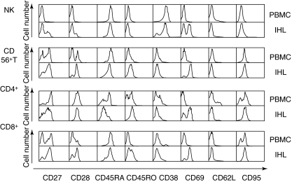
Fig. 3.
All four major intrahepatic lymphocytes (IHL) subsets from the living donor liver displayed increased activation markers, compared to blood cells (peripheral blood mononuclear cells). The fluorescence activated cell sorter histograms show a back-to-back comparison of blood versus liver-derived natural killer, CD56+ T, CD4+ T and CD8+ T cells for eight activation and differentiation markers. See also Tables 1 and 2 for a summary of the data from 10 donors.

