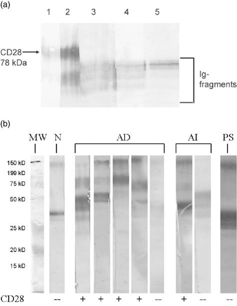
Fig. 1.
(a) Cleavage products after digestion of CD28Ig fusion protein with trypsin. Lane 1, CD28 stained with mouse anti-human CD28 mAb. Lane 2, CD28 stained with biotinylated polyclonal mouse anti-human CD28 antibody. Some smaller fragments of CD28-Ig are detected by this polyclonal antibody but not by the mAb. Lanes 3–5, detection of Ig cleavage products by rabbit anti-human IgG (lane 3), by goat anti-human IgG (lane 4) and by mouse anti-human Fc (lane 5) polyclonal antibodies. (b) Immunoblots of four patients with atopic dermatitis (AD) and autoantibodies to CD28 and one AD patient without CD28 autoantibodies. As an example of CD28 autoantibodies in autoimmune disease (AI) the immunoblot of a patient with scleroderma is presented. The patient with epidermolysis bullosa acquisita (AI) and the patient with psoriasis (PS) were negative for CD28 autoantibodies. Additionally, one immunoblot of a negative serum from a blood donor fromthe control group (N) and the molecular weight standard (MW) is shown.

