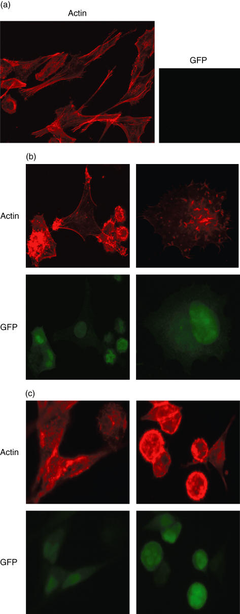
Fig. 1.
Melanoma cells were cultivated on coverslips for 2 days. Cells were infected by vaccinia virus [multiplicity of infection (MOI) 5:1] or left uninfected. Staining for filamentous actin was performed with tetramethylrhodamine-B-isothiocyanate (TRITC)-conjugated phalloidine. Pictures were taken on a Leica fluorescence microscope (DMRB + RD) at low magnification (200 ×) for survey and in detail (up to 1000 ×). (a) Uninfected melanoma cells with a fusiform morphology and strong actin filaments. (b) Western Reserve (WR)-infected melanoma cells: rounded cells, actin clumps and comet-like tails appear. (c) Modified virus Ankara (MVA)-infected melanoma cells: similarly rounded cells, actin clumps and comet-like tails are visible.

