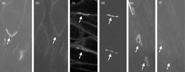
Fig. 5.
Assessment of C9/membrane attack complex (MAC) deposition at the acetylcholine receptor (AChR) in wild-type, C6-deficient and naive Lewis rats. Soleus muscle from naive rats (a,b) and wild-type (c,d) or C6-deficient (e,f) Lewis rats induced for experimental autoimmune MG (EAMG) were harvested, and flash-frozen in isopentane as described. Ten μm thin sections were cut and stained for AChR (a,c,e) using rhodamine-conjugated bungarotoxin. Sections were double-stained for rat C9/MAC (b,d,f) using rabbit anti-rat C9, and detected with anti-rabbit-Ig-fluorescein isothiocyanate (FITC) conjugate. Sections were mounted in VectorShield and analysed under an inverted fluorescent microscope. Images were taken at 800× magnification. Arrows in paired images show identical regions stained in one or both.

