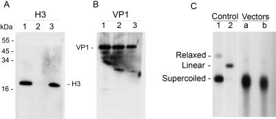
Figure 5. Analysis of the particles.
A,B-Proteins were separated by elecrtrophoresis in Tricine buffer on 16% polyacrylamide gel. Western blotting was performed with polyclonal antibody against histone H3 (Upstate) (A) and against VP1 (B). M–size marker; 1–Nuclear extracts used in the packaging reaction; 2–In vitro assembled particles, fraction 9 of Fig. 4. 3–wild type SV40. C-Analysis of the packaged DNA. Particles purified by ultrafiltration were treated with Tris base (200 mM) in presence of 25 mM EGTA and 25 mM DTT for 1 hr at 37°C. DNA was extracted by phenol-chloroform treatment in presence of 1% SDS, and analyzed by Southern blotting with pGL3-control as a probe.

