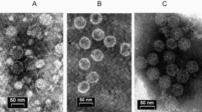
Figure 6. Structure of the nanoparticles.
Transmission electron microscopy pictures of (A) VLPs, (B) in vitro packaged nanoparticles, and (C) wild type SV40. Samples were adsorbed onto formvar-carbon-coated copper grids and stained with 1% sodium phosphotungstate, pH 7.0. The samples were viewed in a Philips CM-12 electron microscope, using a voltage of 100 kV, and photographed at a magnification of 53,000×.

