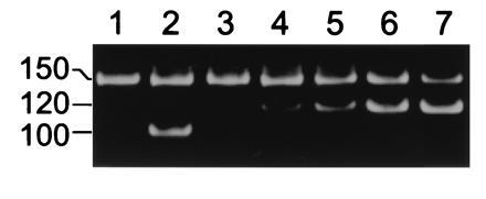
Figure 4.

Single-stranded DNA traversed the α-hemolysin pore. The PCR-amplified product (lane 1) of a 40 μl sample of the trans chamber compared with (lane 2) the similarly amplified product of a 109-fold diluted sample of the cis chamber. All samples were taken following an 8 channel experiment in which a mixture of 150-nt single-stranded DNA (see Materials and Methods) and 100-nt double-stranded DNA was added to the cis chamber. Material in the trans chamber sample reflects accumulation during application of −120 mV (cis chamber negative) for 42 min. Lanes 3-7: example of competitive PCR analysis (25) to show accumulation of single-stranded molecules in the trans chamber. The results show that the 40-μl sample used here contained ≈400 of the 150-nt single-stranded molecules. To each of five 40-μl samples of the trans chamber were mixed (lanes 3–7) 0, 100, 200, 400, or 800 molecules of a competing 120-nt DNA that was PCR amplified using the same primers as the 150-nt DNA. The position of the amplified 150-, 120-, and 100-nt polymers is shown at left. Lanes 1 and 3 show independently amplified 40-μl samples of the same trans chamber.
